
Veterinary Advanced Imaging Advisor · Multislice CT
Choose the clinical tier. Then the exact scanner.
Multislice CT can support fast, versatile full-body imaging across emergency, specialty, referral, and advanced programs. The right direction is defined by the protocols, motion demands, patient range, throughput, facility, and lifetime support—not slice count alone.
Start with the studies the practice must perform.
Before comparing manufacturers, we define the clinical job. Two hospitals can scan similar anatomy and still need different systems because of motion sensitivity, patient size, emergency access, cardiac ambitions, expected volume, team experience, and the way images will be interpreted.
Full-body versatility
Thoracic, abdominal, orthopedic, spinal, neurologic, oncology, and trauma protocols each place different demands on acquisition and reconstruction.
Emergency and throughput
Case arrival patterns, anesthesia time, repeat risk, reconstruction speed, and scanner availability determine whether speed changes the workflow.
Motion and detail
Breathing, heart motion, patient size, metal, contrast timing, and target anatomy help define detector, rotation, software, and tube requirements.
Cardiac CT
Here, cardiac means an intentional advanced cardiac scanning program led by a board-certified veterinary cardiologist—not routine thoracic imaging.
Use slice count as a tier—not a shortcut.
Block Imaging groups CT systems by capability and ownership range. In veterinary medicine, slice count helps frame the comparison, but detector coverage, rotation speed, tube capacity, software, reconstruction, serviceability, and the exact protocols can matter just as much.
General-purpose multislice CT
A strong starting direction for many specialty, emergency, full-body, oncology, orthopedic, spinal, and non-cardiac protocols when volume and motion demands are moderate.
- Lower acquisition and service exposure
- Verify tube capacity for expected volume
- Confirm every required protocol before selection
More speed and advanced flexibility
A direction to evaluate when motion, throughput, cardiac planning, advanced reconstruction, or specialist protocols require performance beyond a routine 16-slice workflow.
- Block identifies 64-slice as a starting cardiac tier
- Validate the exact veterinary cardiac protocol
- Balance performance with lifecycle cost
High-complexity performance
Consider only when advanced cardiac, demanding motion-sensitive studies, broad coverage, or sustained high-volume workflow clearly justify the added system and service complexity.
- Protocol-driven—not prestige-driven
- Higher acquisition and service exposure
- Confirm specialist, facility, and utilization readiness
Cardiac changes the requirement
A higher slice tier is considered when the practice is building advanced cardiac CT with a board-certified veterinary cardiologist, defined protocols, appropriate motion management, and a specialist interpretation plan. Slice count alone does not create a cardiac program; the entire clinical and operational pathway must be ready.
Look beyond the slice label.
Use four practical questions: can the patient fit, can the required anatomy fit inside the imaged field, can the detector cover it efficiently, and can the system acquire it fast enough to control motion?
Specifications matter only inside a protocol.
Ask each provider to demonstrate representative veterinary examinations. Patient size, pitch, anesthesia, breath control, reconstruction, contrast timing, detector coverage, and rotation speed determine whether the result fits your medicine.
Five systems commonly compared in veterinary CT projects.
This comparison deck places the Revolution ACT beside established 16-, 64-, and 128-slice platforms. It is a comparison set—not a ranking or a market-share claim. Exact capability depends on the scanner year, configuration, software, tube history, service plan, and the veterinary protocols you intend to run.

GE BrightSpeed 16
Block includes BrightSpeed among its most-requested refurbished CT systems and describes higher-end BrightSpeed configurations as practical for moderate patient volume. It remains a familiar general-purpose comparison when advanced cardiac imaging is not the program’s core requirement.
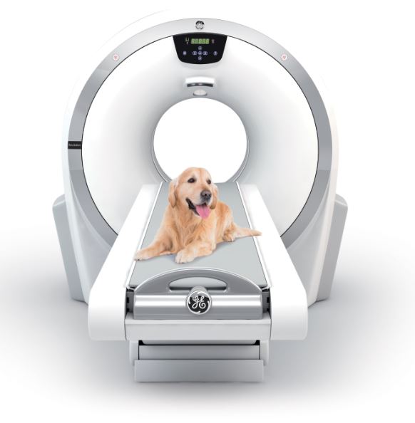
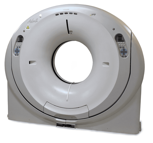
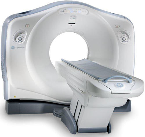

The deck is manual by default. Use the arrows or dots to compare systems, or select Play to advance automatically.
Open the detail you need.
The essentials stay visible above. Use these planning sections when you are ready to compare rooms, manufacturers, service, and the final recommendation method.
Room, power, shielding, and delivery planning
A compact gantry is not the same as a compact finished room. The patient table, service access, egress, anesthesia, console, shielding, HVAC, floor loading, and rigging route all matter.
- BrightSpeed 16: planning references run from roughly 12 ft 10 in × 19 ft 3 in to 13 ft × 20 ft 8 in; 200–480 VAC, three-phase, 90 kVA.
- Revolution ACT: GE’s equipment footprint is not a finished-room requirement; use the exact site drawing for table travel, service, egress, shielding, and 40 kVA input.
- Aquilion 64: about 25–27 m² in reviewed references; 200 VAC, three-phase, 100 kVA.
- LightSpeed VCT 64: roughly 19 ft 2 in × 11 ft 8 in minimum with the smaller table; 380–480 VAC, three-phase, 150 kVA.
- Definition Edge: manufacturer example about 16 ft 9 in × 21 ft 2 in; 400 VAC, 86.5 kVA before configuration-dependent cooling loads.
Before construction, require the exact serial-number-specific site drawing and physicist-approved plan.
Manufacturer families and ownership tradeoffs
- GE: broad refurbished market, parts access, and practical service pathways; verify features and tube on older 16-slice units.
- Siemens: wide performance range and strong advanced options; proprietary tubes and service can raise lifecycle cost.
- Toshiba / Canon: user-friendly, often air-cooled options with a solid tube reputation; confirm local serviceability.
- Philips: strong cardiac capability and durable MRC tube designs; third-party parts and service coverage can be narrower.
Availability changes. Treat these as comparison directions, not a promise that a specific model is in stock.
Service questions that belong in the buying decision
“CT experience” is not specific enough. Document the assigned engineer’s manufacturer and model experience, distance, normal and after-hours response, tube-failure process, parts strategy, preventive maintenance, software coverage, exclusions, and escalation path.
Our multislice service planning uses Block Imaging’s multi-vendor experience so the support pathway is defined before the scanner is finalized.
How the final recommendation is built
The recommendation connects four things: the studies and patient mix; the detector, rotation, tube, reconstruction, and software required for those protocols; the room, power, cooling, shielding, and workflow; and the lifetime budget, training, service, tube exposure, and expected utilization.
You receive the reason for the direction, the closest alternative, what would change the choice, and what still must be verified on the exact quoted system.
Broad selection. Explained recommendation.
Find the multislice system that fits what you are building.
We compare GE, Siemens, Philips, and Toshiba/Canon options against your clinical needs, budget, room, workflow, interpretation plan, and support requirements—then explain why the recommended tier and manufacturer direction make sense.
Source notes: The comparison set uses Block Imaging’s manufacturer and slice-count guide, Siemens Definition comparison, and current Block examples for BrightSpeed and LightSpeed VCT. Technical values were cross-checked against applicable OEM and regulatory documents, but every quoted unit must still be verified by serial number, configuration, software, tube history, room plan, regulatory status, and written service coverage.
Sources: OEM technical literature from GE HealthCare, Siemens Healthineers, Philips, and Canon/Toshiba; Block Imaging product and service resources; and current configuration-specific site-planning documents. Verify the exact model, software level, tube, table, service coverage, and approved site plan.
Veterinary Advanced Imaging Advisor is independently maintained by Chris Weaver, National Imaging Manager at Patterson Veterinary, and is not an official Patterson Companies website. Recommendations begin with clinical need, space, workflow, budget, team readiness, and long-term support. They may include systems Patterson does not sell; when an outside option is the best fit, it will be identified clearly.